
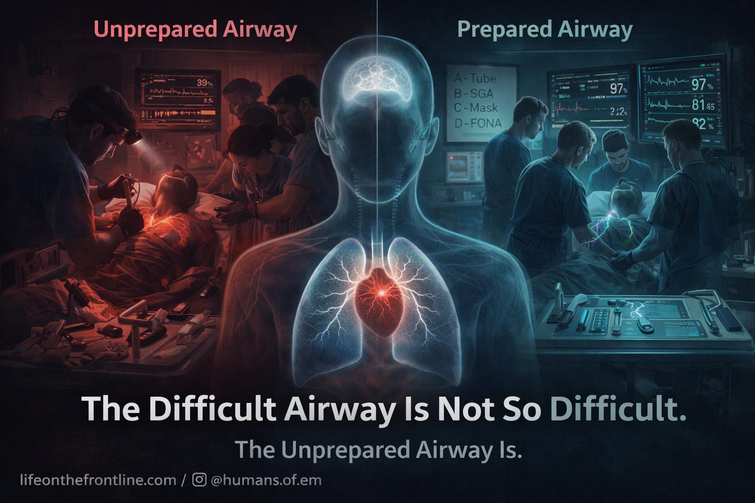
The Difficult Airway Is Not So Difficult. The Unprepared Airway Is.
Integrating physiological and situational airway concepts into emergency practice.

It is 2:17 AM.
The patient is hypoxic. SpO₂ is 82%. Agitated. Diaphoretic.
You push induction.
First attempt fails.
Second attempt fails.
The saturation falls faster than your confidence.
The room gets quieter.
This is the moment that defines airway management.
Not when you intubate.
But when you decide what to do next.
Because modern airway management is not about intubation.
It is about oxygenation.
The Most Important Airway Principle Ever Written
The goal of airway management is not to place a tube.
The goal is to maintain oxygenation.
This distinction seems subtle.
It is not.
It is the difference between controlled progression and catastrophic fixation.
Modern airway guidelines consistently emphasise this shift: prioritising oxygen delivery, limiting attempts, and progressing early through rescue strategies improves outcomes (Ahmad et al., 2025; Myatra et al., 2025).
Hypoxia kills.
Ego does not oxygenate.
Persistence does not oxygenate.
Only oxygenation oxygenates.
The Three Types of Difficult Airway
Every difficult airway belongs to one—or more—of three categories:
Anatomically difficult airway
Physiologically difficult airway
Situationally difficult airway
Understanding this framework changes everything.
Because in emergency medicine, anatomy is rarely the real problem.
Physiology and context are.
The Anatomically Difficult Airway: The Traditional Fear
Defined as difficulty with facemask ventilation, supraglottic airway, laryngoscopy, or surgical airway access (Myatra et al., 2025).
This is the airway we are trained to recognise:
Limited mouth opening
Cervical spine immobility
Facial trauma
Obesity
Airway tumours
This airway is technically difficult.
But often physiologically stable.
These patients can tolerate attempts.
Critically ill patients often cannot.
Won’t discuss much about this.
The Physiologically Difficult Airway: The Airway That Kills
The most dangerous airway is not difficult to intubate.
It is difficult to survive.
The physiologically difficult airway is defined as an airway in which physiological or pathophysiological alterations increase the risk of hypoxaemia, cardiovascular collapse, or death during intubation and transition to positive pressure ventilation (Mosier et al., 2015; Myatra et al., 2022; Ahmad et al., 2025).
You see the cords easily.
You pass the tube easily.
And the patient arrests.
Not because of anatomy.
Because of physiology.
The Five Physiological Phenotypes That Kill Patients
1. Hypoxaemic phenotype
Examples:
ARDS
Severe pneumonia
Pulmonary edema
These patients have no oxygen reserve.
Apnea leads to rapid desaturation.
Peri-oxygenation
— pre , apneic and oxygenation during multiple attempts.
—including head-up positioning, NIV, and high-flow nasal oxygen—prolongs safe apnoea time and reduces hypoxaemia risk (Ahmad et al., 2025; Myatra et al., 2025).
2. Hypotensive / Shock phenotype
Examples:
Septic shock
Hemorrhagic shock
Induction removes sympathetic tone.
Positive pressure ventilation reduces venous return.
Cardiac output collapses.
Cardiovascular collapse during intubation occurs in a substantial proportion of critically ill patients, especially without haemodynamic optimisation (Mosier et al., 2015; Karamchandani et al., 2024).
3. Severe metabolic acidosis phenotype
Examples:
DKA
Septic acidosis
Salicylate poisoning
These patients survive through compensatory hyperventilation.
Apnea removes compensation.
Acidosis worsens.
Cardiac arrest follows.
Preserving spontaneous ventilation may be life-saving.
4. Right ventricular failure phenotype
Examples:
Pulmonary embolism
Severe pulmonary hypertension
Positive pressure ventilation increases pulmonary vascular resistance.
RV fails.
Cardiac output collapses.
5. Neurologic / intracranial hypertension phenotype
Examples:
Severe traumatic brain injury
Intracranial hemorrhage
Even brief hypoxia or hypotension worsens neurologic outcomes.
Attempt to intubation can increase ICP
Physiology must be protected throughout airway management.
The Most Important Question Before Intubation
Not:
Can I intubate this patient?
But:
Can this patient survive intubation?
This single question separates safe airway management from catastrophic airway management.
The Situationally Difficult Airway: When Context Makes the Airway Dangerous
Sometimes anatomy is easy.
Physiology is stable.
But the environment makes the airway dangerous.
This is the situationally difficult airway.
Defined as airway difficulty caused by environmental, logistical, or resource constraints—even when anatomy and physiology are normal (Hung & Murphy, 2010; Karamchandani et al., 2021).
Situational factors that create airway risk
Environment
Emergency department resuscitation
ICU
Prehospital care
Remote locations
Airway management outside controlled environments carries higher complication risk (Lavery & McCloskey, 2008; Karamchandani et al., 2021).
Physical constraints
Confined space
Limited positioning
Poor lighting
These impair visualisation and limit rescue options (Thoeni et al., 2015; Rudolph et al., 2025).
Resource limitations
No videolaryngoscope
Limited assistance
No surgical airway expertise
Operator skill, equipment, and access to help directly influence airway outcomes (Law et al., 2021; Burgess et al., 2022).
Urgency and competing priorities
Active cardiac arrest
Ongoing CPR
Simultaneous resuscitation
These increase cognitive load and reduce airway success.
Human factors contribute significantly to airway complications (Myatra et al., 2025).
The Airway Algorithm That Saves Lives: Plans A, B, C, and D
Airway management is not a single plan.
It is a sequence of oxygenation strategies.
Plan A: Tracheal Intubation
Maximise first-pass success:
Videolaryngoscopy preferred (Ahmad et al., 2025)
Optimal positioning
Neuromuscular blockade
Preoxygenation
First-pass success reduces complications and improves outcomes (Mosier et al., 2015).
Plan B: Supraglottic Airway
Rapid restoration of oxygenation.
Guidelines emphasise switching early between airway devices without fixation on intubation (Myatra et al., 2025).
Plan C: Face Mask Ventilation
Life-saving oxygenation strategy.
Buys time.
Allows physiological stabilisation.
Plan D: Emergency Front-of-Neck Access
Not failure.
Survival.
Emergency cricothyroidotomy is indicated when oxygenation cannot be achieved through other methods (Ahmad et al., 2025; Myatra et al., 2025).
The Vortex Approach: The Cognitive Framework That Prevents Airway Catastrophe
In a true airway crisis, the greatest threat is not anatomy. It is indecision. The Vortex Approach provides a simple, high-stakes cognitive framework designed for these exact moments. Instead of viewing airway management as a rigid sequence, the Vortex conceptualises three non-surgical oxygenation lifelines—facemask ventilation, supraglottic airway, and tracheal intubation—as interchangeable pathways, all aimed at a single goal: effective alveolar oxygen delivery (Nichols et al., 2017; Myatra et al., 2025).
Each attempt should be deliberate and optimised, but fixation on any one technique must be avoided. If oxygenation cannot be achieved despite best efforts in all three lifelines, the Vortex makes the decision clear and immediate: proceed to emergency front-of-neck access.
This framework removes hesitation, reduces cognitive overload, and prioritises oxygenation over procedural persistence. Modern guidelines increasingly emphasise similar principles—early recognition of failure, limiting repeated attempts, and rapid progression to rescue strategies—to prevent hypoxic injury and death (Ahmad et al., 2025; Myatra et al., 2025).
The Vortex does not replace airway skill. It protects decision-making when skill alone is not enough.
The Most Important Intervention Happens Before Induction
Preparation.
Not speed.
Preparation.
Optimise oxygenation.
Optimise hemodynamics.
Prepare equipment.
Prepare backup plans.
Communicate clearly.
Airway strategy must account for anatomy, physiology, environment, operator skill, and resources (Ahmad et al., 2025; Law et al., 2021).
The Most Dangerous Airway Error Is Fixation
One more attempt.
One more try.
Meanwhile, physiology collapses.
Human factors—including fixation and cognitive overload—are major contributors to airway complications (Myatra et al., 2025).
Expert airway clinicians recognise failure early.
They progress deliberately.
They prioritise oxygenation.
The Emergency Physician’s Airway Framework
Before induction, ask:
Do I have Plan A?
Do I have Plan B immediately ready?
Do I have Plan C immediately ready?
Am I prepared for Plan D?
Have I optimised physiology?
Have I accounted for environment?
If the answer is no,
You are not ready.
Not yet.
DIFFICULT AIRWAY & OXYGENATION ALGORITHM
(Emergency / ICU / ED Setting)
Integrates anatomic, physiologic, and situational difficulty with ABCD oxygenation planning.
STEP 1 – DEFINE THE PROBLEM (Before Induction)
Ask three questions:
Anatomy – Is mask / supraglottic / laryngoscopy likely difficult? Look for: limited mouth opening, neck immobility, facial trauma, obesity, tumours, prior difficult airway.
Physiology – Can this patient survive intubation? High‑risk phenotypes: Hypoxaemic (ARDS, pneumonia, pulmonary oedema) Shock / hypotension Severe metabolic acidosis (DKA, sepsis, salicylate) RV failure (PE, pulmonary hypertension) Raised ICP / major brain injury
Situation – Does environment add risk? Location, lighting, space, help, video availability, surgical backup, concurrent CPR, multi‑team resus.
➡️ If any answer is “yes” → treat as difficult airway. Pause and optimise.
STEP 2 – OPTIMISE BEFORE INDUCTION
a. Oxygenation Upright or ramped position. 3–5 min pre‑oxygenation (NIV or HFNO if hypoxaemic). Maintain nasal O₂ / facemask O₂ between attempts.
b. Haemodynamics Correct hypovolaemia, metabolic derangements. Vasopressors on board if hypotensive. Choose induction/paralytic mindful of BP effects.
c. Strategy Define and brief Plan A–D: A – Intubation, B – Supraglottic, C – Mask, D – Front‑of‑neck access.
Team brief: “Two failed attempts or desaturation → move to next plan.”
✔ Proceed only when ready for A, B, C, D and physiology is optimised.
STEP 3 – PLAN A: TRACHEAL INTUBATION (Single Best Attempt) Videolaryngoscopy (preferred), optimal positioning (ear‑to‑sternal notch).
Full NMB unless preserving effort for severe acidosis.
Continue apnoeic oxygenation.
Limit attempt duration; stop if view poor or SpO₂ falls. ❌ If two optimised attempts fail or oxygenation inadequate → move to Plan B/C.
STEP 4 – PLANS B & C: RESCUE OXYGENATION (Vortex Mindset)
Plan B – Supraglottic Airway Insert rapidly after failed intubation. Ventilate to restore SpO₂. Then decide: wake up / continue ventilation / intubate through device.
Plan C – Face‑Mask Ventilation Two‑hand, two‑person technique; airway adjuncts as needed. This is a rescue, not a failure — reassess, re‑oxygenate, re‑plan.
🔁 Cycle between A, B, C only while oxygenation maintained. If can’t oxygenate → go to Plan D immediately.
STEP 5 – PLAN D: EMERGENCY FRONT‑OF‑NECK ACCESS
Trigger: Failure of A + B + C to oxygenate.
Announce: “Can’t oxygenate, can’t ventilate – performing cricothyroidotomy.” Perform scalpel–bougie–tube (or institutional method). Continue efforts above neck while preparing neck.
✅ Plan D is the correct, life‑saving choice – not a failure.
STEP 6 – AFTER SECURING THE AIRWAY
Reassess oxygenation, ventilation, BP, acid–base status. Adjust ventilator to physiology (e.g. ↑PEEP for ARDS, gentle pressures for RV failure, ↑minute ventilation for metabolic acidosis). Quick team debrief: what worked, what to refine next time.
Concept Summary: “Three Difficulties + ABCD Oxygenation = Safe Airway.”
Prepare early, optimise physiology, act decisively, and move forward when oxygenation fails.
Final Thought
The most dangerous airway is not the one you cannot see.
It is the one whose physiology you do not respect.
The best airway clinicians do not just secure the airway.
They protect oxygenation.
They protect physiology.
They protect life.
Because the difficult airway is not difficult.
The unprepared airway is.
References :
Ahmad I, El-Boghdadly K, Bhagrath R, et al. Difficult Airway Society guidelines for awake tracheal intubation in adults. Anaesthesia. 2025;80(3):395–418.
Myatra SN, Shah A, Kundra P, et al. All India Difficult Airway Association 2025 guidelines for management of difficult airway in adults. Indian J Anaesth. 2025;69(2):89–112.
Mosier JM, Joshi R, Hypes CD, Pacheco GS, Valenzuela TD, Sakles JC. The physiologically difficult airway. West J Emerg Med. 2015;16(7):1109–1117. doi:10.5811/westjem.2015.8.27467
Myatra SN, Divatia JV, Brewster DJ. The physiologically difficult airway: an emerging concept. Curr Opin Anaesthesiol. 2022;35(2):115–121. doi:10.1097/ACO.0000000000001102
Karamchandani K, Nasa P, Jarzebowski M, et al. Tracheal intubation in critically ill adults with a physiologically difficult airway: an international Delphi consensus study. Intensive Care Med. 2024;50(11):1563–1579. doi:10.1007/s00134-024-07578-2
Kornas RL, Owyang CG, Sakles JC, Foley LJ, Mosier JM. Evaluation and management of the physiologically difficult airway: consensus recommendations. Anesth Analg. 2020;132(2):395–405. doi:10.1213/ANE.0000000000005233
Pacheco GS, Hurst NB, Patanwala AE, et al. First-pass success without adverse events is reduced equally with anatomically and physiologically difficult airways. West J Emerg Med. 2021;22(2):360–368. doi:10.5811/westjem.2020.10.48887
Jabaley CS, Groff RF, Sharp WW, et al. Managing the physiologically difficult airway in critically ill adults. Crit Care. 2023;27(1):1–12. doi:10.1186/s13054-023-04371-3
Law JA, Duggan LV, Asselin M, et al. Canadian Airway Focus Group updated consensus-based recommendations for management of the difficult airway: part 1 and part 2. Can J Anaesth. 2021;68(9):1373–1436. doi:10.1007/s12630-021-02007-0
Hung O, Murphy MF. Context-sensitive airway management. Anesth Analg. 2010;110(4):982–983. doi:10.1213/ANE.0b013e3181d48bbb
Burgess MR, Schauer SG, De Lorenzo RA. The difficult airway redefined. Prehosp Disaster Med. 2022;37(6):723–726. doi:10.1017/S1049023X22001455
Lavery GG, McCloskey BV. The difficult airway in adult critical care. Crit Care Med. 2008;36(7):2163–2173. doi:10.1097/CCM.0b013e31817d7ae1
Thoeni N, Piegeler T, Brueesch M, et al. Incidence of difficult airway situations during prehospital airway management. Resuscitation. 2015;90:42–45. doi:10.1016/j.resuscitation.2015.02.010
Rudolph SS, Root C, Tvede MF, et al. Confined space airway management: a narrative review. Scand J Trauma Resusc Emerg Med. 2025;33(1):57. doi:10.1186/s13049-025-01357-8
Liaqat T, Amjad M, Cherian SV. Difficult airway management in the intensive care unit: a narrative review. J Clin Med. 2025;14(14):4930. doi:10.3390/jcm14144930
Nichols D, Kelly FE, Cook TM. The Vortex approach: a universal high-acuity implementation tool for emergency airway management. Anaesthesia. 2017;72(9):1063–1069. doi:10.1111/anae.13967
Dunford B, Sutterfield B, Roberts W, et al. Difficult airway management: analysis of systematic review evidence underpinning clinical practice guidelines. Anaesth Crit Care Pain Med. 2025;44:101534. doi:10.1016/j.accpm.2025.101534
Cabrini L, Landoni G, Baiardo Redaelli M, et al. Tracheal intubation in critically ill patients: a systematic review. Crit Care. 2018;22(1):6.
Mosier JM. Physiologically difficult airway and management considerations. Curr Anesthesiol Rep. 2024;14(4):446–457.
Jung H. A comprehensive review of difficult airway management strategies for patient safety. Anesth Pain Med. 2023;18(4):331–339.