

By-
Dr Arihant Jain, MD | lifeonthefrontline.com
Instagram: @humans.of.em
X | Linkedin | ORCID
————————————————————————
A patient arrives in the ED with severe dyspnoea. Bibasal crackles. B-lines on lung ultrasound. A raised NT-proBNP. Perhaps some peripheral oedema.
The reflex is familiar:
“This is acute heart failure. Give IV Lasix.”
But the 2026 ESC Guidelines for the management of heart failure encourage a more nuanced approach. The question is not simply:
“Does this patient have heart failure?”
Nor even:
“How much furosemide should I give?”
The better questions are:
What is the clinical phenotype?
Where is the congestion?
Is the patient perfused?
Is this true volume overload or predominantly fluid redistribution?
And, after treatment, how will I know that decongestion is actually occurring?
For every physicians, this may be one of the most useful ways to translate the 2026 ESC update into bedside practice.
1. First, the terminology has changed
The 2026 ESC Guidelines have eliminated the previous HFmrEF category.
The new classification is:
HFrEF: LVEF <50% with symptoms and/or signs of HF
HFpEF: LVEF ≥50% with symptoms/signs of HF plus objective evidence of structural and/or functional abnormalities consistent with LV diastolic dysfunction or raised LV filling pressures, supported by elevated natriuretic peptides.
The guideline has also replaced the term “acute heart failure” with “decompensated heart failure (DHF)”.
For the acute care physician, however, the more important change is conceptual:
DHF is not one phenotype, and therefore it should not have one reflex treatment.
2. Start with phenotype, not the prescription pad
The 2026 ESC framework separates patients into clinically important phenotypes:
Cardiogenic shock
Acute pulmonary oedema
Decompensated left-sided HF
Decompensated right-sided HF
And these phenotypes can overlap.
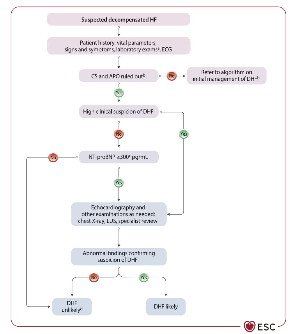
Figure 1. Diagnosing decompensated heart failure. Source: 2026 ESC Guidelines for the management of heart failure.
The guideline also provides non-age-adjusted natriuretic peptide rule-out thresholds for DHF:
NT-proBNP <300 pg/mL
BNP <100 pg/mL
MR-proANP <120 pg/mL
Age-related NT-proBNP thresholds can then support a likely diagnosis in older patients.But once DHF is suspected, the next question is:
What phenotype am I treating?
3. Congestion and perfusion: the two questions that matter
The guideline provides a useful separation between left-sided congestion, right-sided congestion and hypoperfusion.
Left-sided congestion
Think:
Dyspnoea
Orthopnoea
Cough
Tachypnoea
Rales
S3
Pleural effusion
Elevated natriuretic peptides
Right-sided congestion
Think:
Peripheral oedema
Abdominal distension
Hepatomegaly
Raised JVP
Hepatojugular reflux
Pleural effusion
Hypoperfusion
Think:
Cold/sweaty extremities
Pale skin
Dizziness
Confusion
Oliguria
Narrow pulse pressure
Elevated lactate
AKI
Hepatic injury.
So at the bedside, I would simplify it to:
WET or DRY?
WARM or COLD?
LEFT, RIGHT or BOTH?
That is more useful than simply labelling the patient “acute HF.”
4. Not every “wet” patient needs aggressive Lasix
This is perhaps the most important message for acute care physicians.
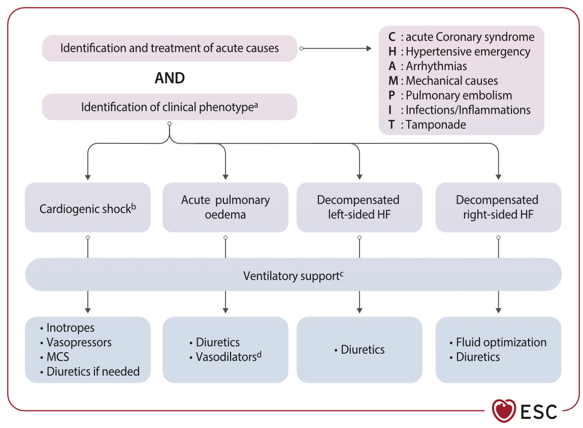
Figure 2. Initial management of decompensated heart failure. Source: 2026 ESC Guidelines for the management of heart failure.
The hypertensive pulmonary oedema patient is the classic example
Consider:
BP 200/110 mmHg
Severe respiratory distress
Diffuse B-lines
Orthopnoea
It is tempting to interpret this as:
“Massive fluid overload → massive dose of furosemide.”
But the guideline specifically highlights that acute pulmonary oedema can occur predominantly because of fluid redistribution into the pulmonary circulation, rather than massive total-body volume overload.
These patients usually do not need high doses of diuretics and may instead require vasodilator therapy. The guideline allows IV vasodilators to be considered as initial therapy in DHF when SBP >110 mmHg, particularly to improve symptoms and reduce congestion.
So:
Pulmonary congestion ≠ automatically massive volume overload.
And:
B-lines ≠ a prescription for high-dose Lasix.
The clinical context matters.
5. Right-sided failure is another phenotype where “Lasix first” deserves caution
Decompensated isolated right-sided HF is characterized by elevated right-sided pressures, systemic congestion and, in advanced disease, hypoperfusion.
The guideline specifically highlights the importance of RV function, ventricular interdependence and the effects on kidney and liver function. Pulmonary embolism and RV infarction may also need to be excluded as acute causes. So a patient with:
Raised JVP + oedema + hepatomegaly + hypotension
is not simply a “fluid overloaded patient.”
The question becomes:
Is this a congested patient who needs decongestion, or a preload-dependent/hypoperfused patient whose haemodynamics need stabilization first?
That distinction is critical.
6. If the patient IS congested, then decongestion becomes the goal
For the genuinely congested patient, IV loop diuretics remain the cornerstone of treatment.
—> For a diuretic-naïve patient:
40 mg IV furosemide or equivalent
—> For a patient already receiving oral loop diuretics:
approximately twice the usual daily oral loop-diuretic dose IV can be considered.
But here’s where the approach becomes much more interesting.
The dose is not the endpoint. The response is.
7. Don’t just give Lasix. Test the response.
Historically, the question after giving furosemide was often:
“Did the patient pee?”
The 2026 ESC approach is more objective.
At approximately 2 hours:
Spot urinary sodium ≥70 mEq/L
OR
During the first 6 hours:
Urine output ≥100 mL/hour
These are the guideline’s markers of a satisfactory early diuretic response.
This gives us a very useful bedside concept:
Don’t ask only how much diuretic you gave.
Ask how much sodium and water the patient actually removed.
The PUSH-AHF and ENACT-HF studies showed that uNa-guided strategies can increase natriuresis/diuresis, although importantly, no trial has yet demonstrated a mortality or recurrent-HF-hospitalization benefit from uNa-guided therapy itself.
So uNa should be viewed as a response-monitoring tool, not a magic prognostic marker.
8. What if the patient doesn’t respond?
This is where the guideline becomes particularly practical.
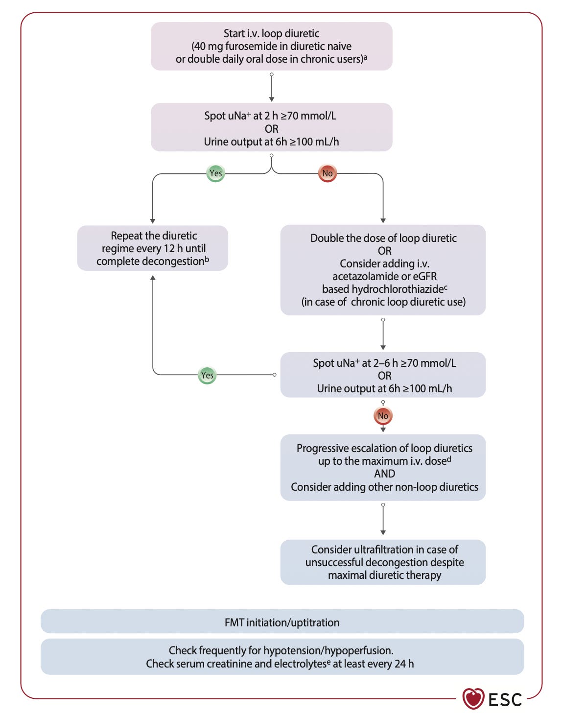
Figure 15. Management of decongestion. Source: 2026 ESC Guidelines for the management of heart failure.
If congestion persists, progressively escalate pharmacological decongestion and consider sequential nephron blockade. In refractory cases despite maximal pharmacological therapy, ultrafiltration may be considered.
9. Sequential nephron blockade: don’t just keep pushing the loop
The guideline incorporates acetazolamide and thiazide-type therapy into the strategy for inadequate diuretic response. In ADVOR, adding IV acetazolamide 500 mg daily to loop diuretics increased successful decongestion, defined as absence of signs of volume overload within three days.
Hydrochlorothiazide increased weight loss and 24-hour diuresis in CLOROTIC, but was associated with more worsening kidney function and hypokalaemia and did not improve patient-reported dyspnoea. Neither trial demonstrated benefit on hard clinical endpoints.
This is an important distinction:
More diuresis is not automatically better medicine.
The objective remains:
Effective, safe decongestion. Not maximum urine output at any cost.
10. The creatinine trap
One of the commonest reasons we stop effective decongestion is:
“Creatinine has gone up.”
But the ESC guideline specifically cautions against interpreting kidney function in isolation. Small, transient rises in serum creatinine during diuresis are not associated with poor outcomes when adequate decongestion is achieved.
Therefore:
Creatinine ↑ + congestion improving + perfusion adequate
does not automatically mean: STOP DIURESIS.
Compare that with:
Creatinine ↑ + persistent congestion + hypotension/hypoperfusion + poor diuretic response
Now we have a completely different problem. The kidney number needs to be interpreted alongside:
Congestion + perfusion + BP + urine output + natriuresis + trajectory.
11. How congested is the patient?
This is where the concept of a congestion score becomes useful.
The ESC clinical congestion score considers:
Dyspnoea
Orthopnoea
Fatigue
Rales
Oedema
JVP distension
A score of 0 represents absence of clinical congestion, while 1–2 represents mild congestion.
The key is not necessarily the number itself.
It is the trend.
On arrival, After treatment, Before discharge:
Is congestion actually gone?
That is much more meaningful than:
“He made 2.5 litres of urine.”
12. Decongestion is multimodal
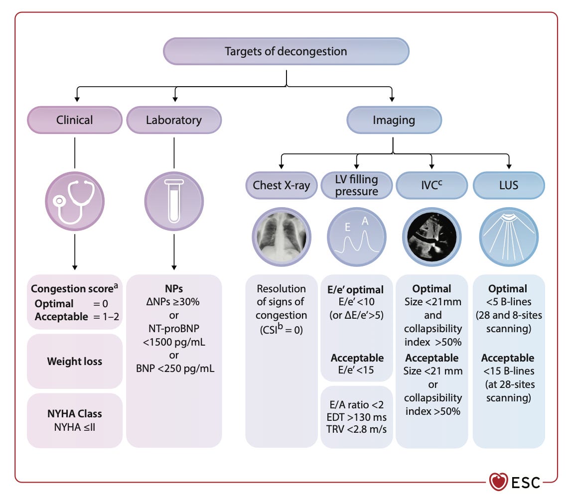
Figure 4. Tools used for assessment of decongestion during the pre-discharge phase. Source: 2026 ESC Guidelines for the management of heart failure.
This is an excellent figure for acute care physicians because it brings together several domains.
Clinical
Congestion score
Weight loss
NYHA class
Laboratory
BNP
NT-proBNP
Imaging
Chest X-ray / congestion score index
LV filling pressure parameters
IVC
Lung ultrasound
The guideline recommends assessment of residual congestion before discharge using clinical, laboratory and imaging techniques.
But there is an important caveat:
These tools are optional, because randomized trial evidence demonstrating improved outcomes from their use is lacking.
So POCUS is not a replacement for examination . It is another piece of the puzzle.
13. The POCUS question should change
Instead of simply asking:
“Does this patient have B-lines?”
ask:
“Where is the congestion, and is it resolving?”
Lung ultrasound
Are there B-lines?
Are they diffuse?
Are they changing with treatment?
Heart
What does LV/RV function look like?
Venous system
Is there evidence supporting systemic venous congestion?
Pleura
Is there an effusion?
And then integrate this with:
JVP + oedema + respiratory examination + urine output + uNa + renal function + BP + perfusion.
The guideline itself includes LUS and other imaging modalities as components of decongestion assessment rather than prescribing one imaging measurement as definitive.
14. “Dry” is not the same as “better”
One of the most important messages in the 2026 guideline is residual congestion.
A patient can:
breathe better,
have less oedema,
produce several litres of urine,
and still be congested. Residual congestion is associated with poor outcomes and increased risk of rehospitalization. Therefore, the guideline recommends careful evaluation before discharge to exclude persistent congestion.
This changes our discharge question.
Not:
“Does the patient feel better?”
But:
“Have we achieved adequate decongestion?”
15. Decongestion is only one phase of the hospitalization
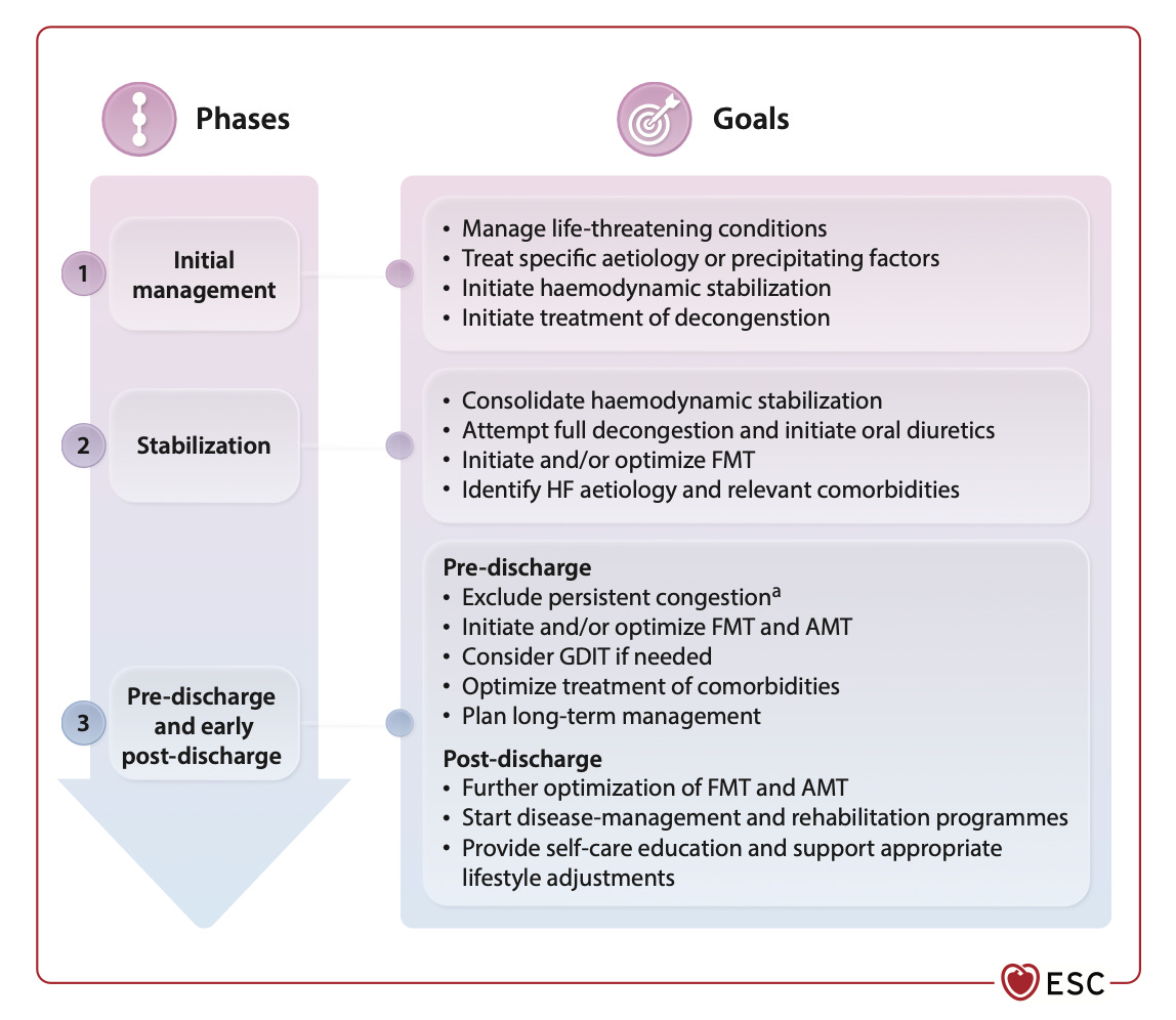
Figure 5. Phases and goals for in-hospital management of decompensated heart failure. Source: 2026 ESC Guidelines for the management of heart failure.
The ESC framework divides inpatient management into three broad phases:
Phase 1 — Initial management
Treat life-threatening conditions
Identify and treat precipitating factors
Stabilize haemodynamics
Initiate treatment of decompensation
Phase 2 — Stabilization
Consolidate haemodynamic stability
Attempt full decongestion
Initiate/optimize foundational medical therapy
Identify and manage comorbidities
Phase 3 — Pre-discharge and early post-discharge
Exclude persistent congestion
Optimize FMT
Determine need for additional/interventional therapies
Plan long-term management.
This is an important departure from the old mental model:
“Treat the acute episode and then deal with chronic HF later.”
16. Start disease-modifying therapy during the hospitalization
The guideline emphasizes that decongestion and foundational medical therapy should proceed in parallel once the patient is stabilized. MRAs, SGLT2 inhibitors and ARNIs can be initiated during the stabilization phase, and the traditional sequential approach to FMT is considered time-consuming; simultaneous implementation is preferred when feasible, with more conservative approaches for selected high-risk patients.
In-hospital initiation of an SGLT2 inhibitor after initial stabilization is recommended to improve quality of life/congestion symptoms and reduce HF hospitalization. And importantly, if a patient was already receiving foundational therapy, discontinuation is not recommended unless there are clear signs of hypoperfusion or another specific clinical indication.
17. The patient with shock is a different conversation
A congested patient who is also hypoperfused cannot simply be treated using the same algorithm as an uncomplicated “warm and wet” patient. The guideline emphasizes that cardiogenic shock is defined by critical end-organ hypoperfusion, and there is no single blood-pressure threshold that defines it. Hypoperfusion can occur even with normal blood pressure.
Look for:
Cold extremities
Confusion
Oliguria
Narrow pulse pressure
Lactate elevation
AKI/hepatic injury
The guideline uses the SCAI shock stages from:
A — At risk
B — Beginning/pre-shock
C — Classic shock
D — Deteriorating
E — Extremis.
And when cardiogenic shock with potential need for temporary mechanical circulatory support is suspected, consultation with a Shock Team is recommended.
18. The bedside algorithm I want to remember
If I had to reduce the 2026 ESC approach to one ED mental model:
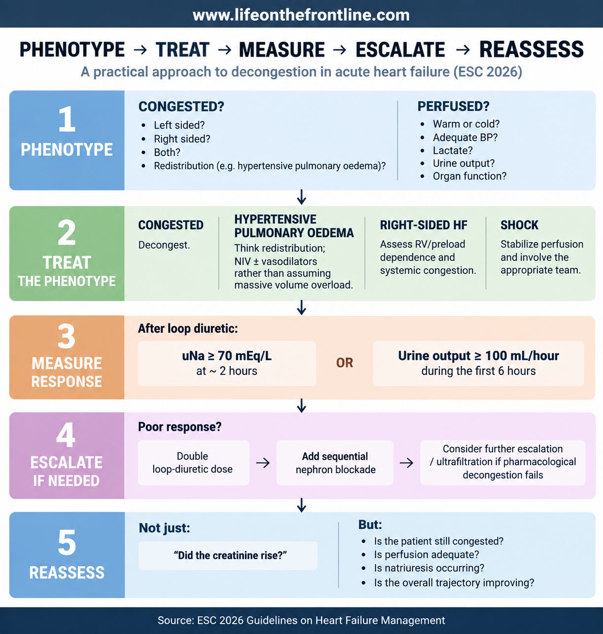
The bigger mindset shift
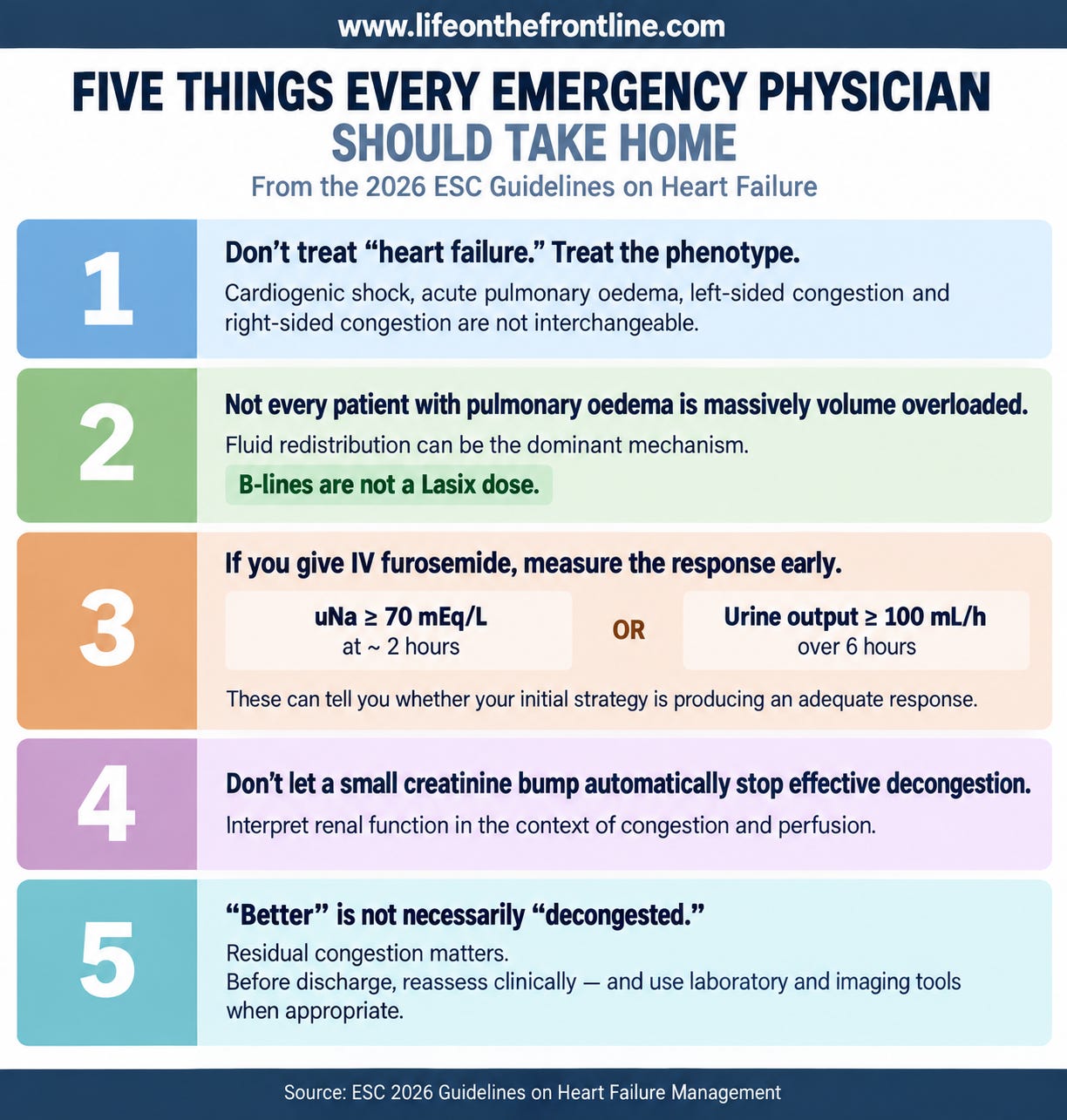
Lasix is a tool. Decongestion is the goal. Phenotyping determines the strategy. And on the frontline, that distinction matters.
Must Read : ESC 2026 Heart Failure Guidelines