

By-
Dr Arihant Jain, MD | lifeonthefrontline.com
Instagram: @humans.of.em
X | Linkedin | ORCID
————————————————————————
We often look at an ABG, see a low PaO₂, and immediately say:
“The patient is hypoxic.”
But technically, that may not be correct.
Hypoxemia and hypoxia are related—but they are not the same thing.
And when reading an ABG, PaO₂ is only the beginning of the oxygenation story.
This is ABG Series #2 — a practical approach to understanding oxygenation before we dive into acid–base disorders.
1. Hypoxemia vs Hypoxia
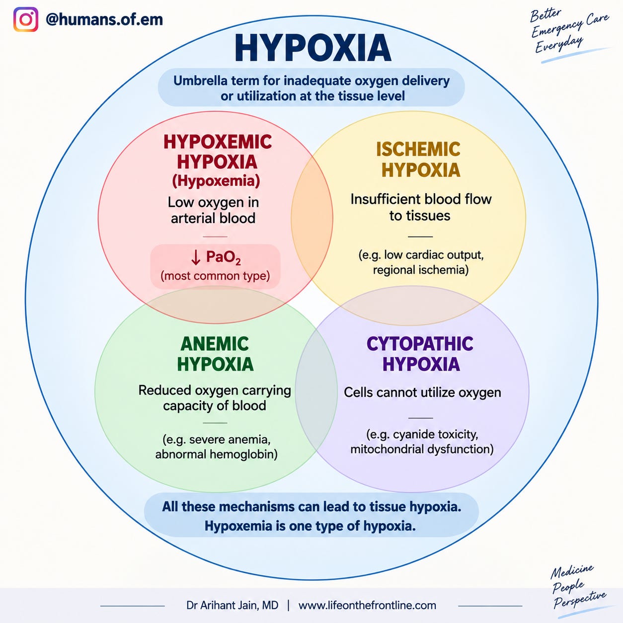
1.1. Hypoxemia
Low oxygen in arterial blood.
On an ABG, we primarily assess this using:
PaO₂
SaO₂
A typical PaO₂ of 70–100 mmHg and SaO₂ of 93–98% as reference values at room air.
1.2. Hypoxia
Inadequate oxygen delivery or utilization at the tissue level.
This is a broader physiological problem.
A patient can therefore have:
Hypoxemia → impaired oxygen delivery → tissue hypoxia
…but tissue hypoxia can also occur without significant hypoxemia.
The infographic above shows four broad mechanisms of hypoxia: hypoxemic, ischemic, anemic and cytopathic hypoxia.
Think of it this way:
Hypoxemia = problem with oxygen in arterial blood
Hypoxia = problem with oxygen availability/use at the tissue level
2. So what exactly does the PaO₂ tell me?
PaO₂ = partial pressure of oxygen dissolved in arterial plasma.
It tells us about the oxygen tension in arterial blood.
It does not directly tell us:
how much oxygen is carried by haemoglobin
whether tissues are receiving enough oxygen
whether cardiac output is adequate
whether cells can utilize oxygen
That’s why PaO₂ should not be interpreted in isolation.
The ABG itself is designed to assess oxygenation, ventilation and acid–base status, while also providing information relevant to oxygen-carrying capacity.
3. The oxygenation question I ask first
When I see an ABG, don’t simply ask:
“Is PaO₂ low?”
Ask:
“Is this PaO₂ appropriate for the amount of oxygen the patient is receiving?”
That’s a much more useful question.
Because:
PaO₂ 80 mmHg on room air
is very different from
PaO₂ 80 mmHg on FiO₂ 0.80.
The number is identical.
The physiology isn’t.
4. Steps to decipher oxygenation status
4.1. Step 1 — Know the FiO₂
Before interpreting PaO₂, determine:
What is the patient breathing?
Room air:
FiO₂ ≈ 0.21
Supplemental oxygen:
FiO₂ depends on the device, flow, patient breathing pattern and interface.
This is particularly important because calculations such as the A–a gradient require an accurate FiO₂; estimation becomes less reliable with devices such as nasal cannulae , simple face masks and NRBM.
4.2. Step 2 — Look at PaO₂ and SaO₂
A “normal” PaO₂ doesn’t automatically mean normal oxygenation.
Always interpret it in relation to FiO₂.
4.3. Step 3 — Calculate the P/F ratio
One of the simplest ways to account for the inspired oxygen is:
P/F ratio = PaO₂ ÷ FiO₂
Example:
PaO₂ = 60 mmHg
FiO₂ = 0.50
Therefore:
P/F = 60 ÷ 0.5 = 120 mmHg
The source material describes a normal P/F ratio as approximately 300–500 mmHg, with lower values indicating impaired gas exchange.
Why is this useful?
Because:
PaO₂ tells you the oxygen tension.
P/F tells you how effectively the lungs are achieving that PaO₂ given the oxygen being supplied.
A simple bedside comparison
Patient A
PaO₂ = 80
FiO₂ = 0.21
P/F ≈ 381
Reasonable oxygenation.
Patient B
PaO₂ = 80
FiO₂ = 0.80
P/F = 100
Very different story.
Same PaO₂.
Completely different oxygenation efficiency.
That’s why:
Never interpret PaO₂ without looking at FiO₂.
4.4. Step 4 — When you need to know why the PaO₂ is low: A–a gradient
The alveolar–arterial oxygen gradient compares:
Oxygen in the alveoli with Oxygen actually reaching arterial blood.
A–a gradient = PAO₂ − PaO₂
The A–a gradient as a way of assessing the efficiency of pulmonary oxygen transfer and differentiating pulmonary from extrapulmonary causes of hypoxemia.
How do we calculate PAO₂?
Using the alveolar gas equation:
PAO₂ = FiO₂ × (Patm − PH₂O) − PaCO₂/R
At sea level:
Patm ≈ 760 mmHg
PH₂O ≈ 47 mmHg
R ≈ 0.8
So on room air:
PAO₂ ≈ 0.21 × (760 − 47) − PaCO₂/0.8
The ABG material provides this equation and notes that the respiratory quotient is approximately 0.8 under steady-state conditions.
The A–a gradient gives you a physiological clue
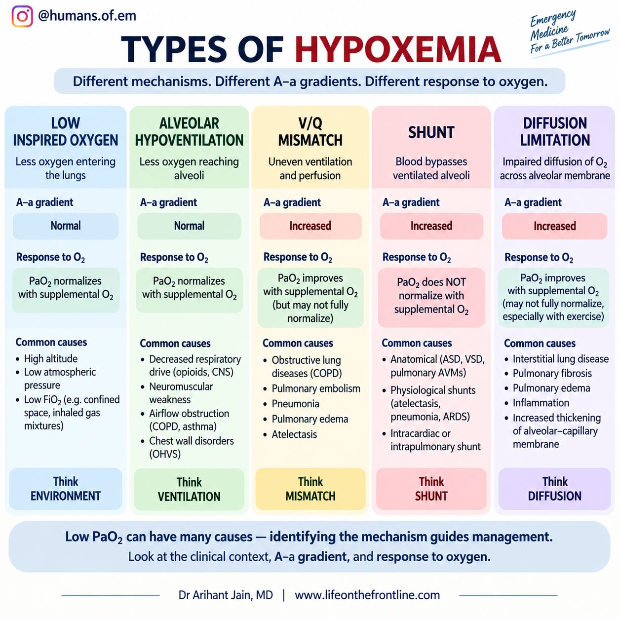
Normal/near-normal A–a gradient
Think:
The lungs are transferring oxygen reasonably well.
Possible causes of hypoxemia include:
low inspired oxygen
hypoventilation
Increased A–a gradient
Think:
There is a problem with pulmonary oxygen transfer.
Common mechanisms include:
V/Q mismatch
shunt
diffusion limitation
The infographic illustrates these mechanisms and shows that V/Q mismatch, shunt and diffusion limitation produce an increased A–a gradient, whereas low inspired oxygen and hypoventilation can have a relatively preserved gradient.
5. But here’s where hypoxemia ≠ hypoxia becomes important
Imagine a patient with:
Severe anaemia
PaO₂ → normal
SaO₂ → normal
Yet the patient may have impaired oxygen delivery because there isn’t enough haemoglobin available to carry oxygen.
Similarly, a patient with:
Low cardiac output
may have adequate arterial oxygenation but inadequate oxygen delivery to tissues.
And in certain toxic states, cells may be unable to utilize oxygen despite adequate arterial oxygen.
So:
Normal PaO₂ does not guarantee normal tissue oxygenation.
6. The bigger equation: Oxygen delivery
Ultimately, tissue oxygen delivery depends on:
DO₂ = Cardiac Output × CaO₂
And arterial oxygen content:
CaO₂ ≈ (1.34 × Hb × SaO₂) + (0.003 × PaO₂)
The important takeaway is that most oxygen content is carried by haemoglobin, while only a very small amount is dissolved in plasma. The ABG infographic in the source explicitly illustrates this relationship.
So a useful mental model is:
PaO₂
→ oxygen tension
SaO₂
→ percentage of Hb binding sites occupied
Hb
→ how much oxygen-carrying protein is available
Cardiac output
→ how much oxygenated blood reaches tissues
Cellular utilization
→ whether tissues can actually use the delivered oxygen
7. My bedside oxygenation checklist
When I open an ABG, I would go:
1. Is it actually arterial?
2. What is the FiO₂?
3. What is the PaO₂?
4. What is the SaO₂?
5. Is the PaO₂ appropriate for the FiO₂?
6. Calculate P/F ratio when clinically useful
PaO₂ ÷ FiO₂
7. If hypoxemia is present, ask:
Is the A–a gradient increased?
8. Finally ask the bigger question:
Is this only hypoxemia—or is the patient actually suffering from impaired oxygen delivery?
The one-minute takeaway
1. HYPOXEMIA
⬇️
Low arterial oxygen
Think: PaO₂ / SaO₂
↓
2. HYPOXIA
⬇️
Inadequate oxygen availability or utilization at the tissue level
Think: DO₂ = CO × CaO₂
↓
3. ABG oxygenation
Don’t stop at PaO₂.
PaO₂ → FiO₂ → P/F ratio → A–a gradient → oxygen content → tissue oxygen delivery
ABG Series #1 — The take-home message
A low PaO₂ tells you that the blood is hypoxemic.
It doesn’t, by itself, tell you whether the tissues are hypoxic.
And conversely:
Normal PaO₂ doesn’t guarantee adequate tissue oxygen delivery.
Read the ABG.
Then read the physiology.