

By-
Dr Arihant Jain, MD | lifeonthefrontline.com
Instagram: @humans.of.em
X | Linkedin | ORCID
————————————————————————
Hyponatremia is one of the most common electrolyte abnormalities encountered in acute care. Yet when the sodium comes back at 118 mmol/L, the first instinct is often:
“How do I increase the sodium?”
That is not the first question we should ask.
Questions are:
Is the patient symptomatic?
Is this truly hypotonic hyponatremia?
How quickly did it develop?
And what is driving it?

Once those questions are answered, treatment becomes much easier. The following is a practical approach to hyponatremia for the ED and acute-care setting.
Step 1: Is the patient symptomatic?
This is the first and most important question.
Hyponatremia can range from completely asymptomatic to severe neurological deterioration.
Severe symptoms → treat as a neurological emergency
Think “cerebral edema / impending brain injury.”
Seizures
Marked reduction in consciousness
Coma / GCS ≤8
Deep or abnormal somnolence
Persistent vomiting
Cardiorespiratory distress/arrest
Features suggesting raised ICP, such as bradycardia + hypertension + abnormal respiration, may occur in extreme cases
Severe neurological symptoms suggest clinically significant cerebral edema. Acute or severe symptomatic hyponatremia is a medical emergency. Do not wait for the complete diagnostic work-up before treating the cerebral edema.
US vs European Guidelines: How Should We Treat Symptomatic Hyponatremia?
Both the US and European recommendations consider acute or severe symptomatic hyponatremia a medical emergency and recommend 3% hypertonic saline as the treatment of choice. The important principle is that treatment is aimed at rapidly reversing cerebral edema and improving neurological symptoms but not normalizing the serum sodium concentration. The two approaches are broadly similar but differ in the dose and delivery of hypertonic saline.
The US recommendations suggest 100 mL of 3% saline over 10 minutes, which may be repeated up to three times, with an initial goal of increasing serum sodium by approximately 4–6 mmol/L. The European guideline instead recommends 150 mL of 3% saline over 20 minutes, with sodium reassessed after 20 minutes and the bolus repeated if necessary, up to three times, aiming for approximately a 5 mmol/L increase during the first hour or until symptoms improve.
The difference becomes more interesting when symptoms are moderate rather than severe. The European guideline recommends a single 150-mL bolus of 3% saline over 20 minutes, while the US recommendations describe a more controlled continuous infusion of 3% saline at 0.5–2 mL/kg/hour. The European approach emphasizes that moderately symptomatic patients should undergo prompt diagnostic assessment and cause-specific treatment, with a relatively modest sodium rise targeted over 24 hours.
The two guidelines also differ somewhat in how they approach the rate of correction. The European guideline recommends limiting the sodium increase to 10 mmol/L during the first 24 hours and an additional 8 mmol/L during each subsequent 24-hour period. The US recommendations are somewhat more conservative in their suggested correction target, generally aiming for 4–8 mmol/L/day, while allowing a maximum of approximately 10–12 mmol/L/day in patients without major risk factors for osmotic demyelination. In patients at particularly high risk of ODS such as those with alcohol excess, malnutrition, hypokalemia, or advanced liver disease the US recommendations advocate an even more conservative approach, with a maximum correction of approximately 4–8 mmol/L/day. Importantly, these numbers should be understood as upper limits, not therapeutic targets.
A practical way to reconcile the two approaches at the bedside is therefore: if the patient has severe neurological symptoms, do not delay treatment while completing the diagnostic work-up—give 3% saline as a monitored bolus and reassess both symptoms and sodium frequently. Once neurological symptoms improve after a modest rise in sodium, stop hypertonic saline and move toward cause-specific treatment. If symptoms fail to improve, reconsider whether hyponatremia is actually responsible for the neurological presentation and continue treatment only with close biochemical monitoring. The European guideline specifically recommends stopping hypertonic saline once symptoms improve after an approximately 5 mmol/L rise, while continuing therapy and reassessing alternative causes if symptoms persist.
Finally, both approaches place major emphasis on avoiding overcorrection. If the sodium begins rising faster than intended, treatment should be interrupted and free-water losses replaced; desmopressin can be used to stop ongoing water diuresis, with hypotonic fluid such as 5% dextrose used when relowering is required. The attached review emphasizes that the combination of desmopressin and free-water replacement can both prevent further sodium rise and, when necessary, actively relower the sodium toward the intended target.
The bedside takeaway
US: 100 mL 3% saline → 10 min → repeat as needed → aim for ~4–6 mmol/L rise.
Europe: 150 mL 3% saline → 20 min → repeat as needed → aim for ~5 mmol/L rise in the first hour.
Both: Treat the neurological emergency, reassess frequently, and never mistake the correction limit for the correction target.
Step 2: How quickly did the sodium fall?
The brain adapts to chronic hyponatremia.
That means the speed of onset matters at least as much as the absolute sodium value.
Acute
<48 hours
Chronic
>48 hours
Unknown duration?
For practical purposes, treat it as chronic when you cannot establish that it is acute.
Why?
Because a chronically hyponatremic brain has adapted to the low osmolality. Rapidly increasing plasma osmolality can injure the adapted brain and cause osmotic demyelination syndrome (ODS).
So remember:
A sodium of 118 is not automatically an indication to rapidly normalize the sodium.
The clinical context matters.
Step 3: Is this actually hypotonic hyponatremia?
A low serum sodium does not always mean that the plasma is hypotonic.
This is where serum osmolality becomes useful.
Low sodium + LOW serum osmolality
→ Hypotonic hyponatremia (True Hyponatremia)
This is the major group we need to investigate further.
Low sodium + NORMAL osmolality
→ Think pseudo-hyponatremia
Hyperproteinemia
Multiple myeloma
Waldenström macroglobulinemia
Marked hypergammaglobulinemia
Hyperlipidemia
Severe hypertriglyceridemia
Occasionally severe hypercholesterolemia
Low sodium + HIGH osmolality
→ Think hypertonic hyponatremia
A classic example is severe hyperglycemia, where glucose contributes to plasma osmolality and lowers the measured sodium concentration.
So:
Do not start by asking “Why is the sodium low?”
First ask: “Is this true hypotonic hyponatremia?”
Step 4: If it is hypotonic — what is the kidney doing?
This is where the diagnostic approach becomes much simpler.
Send:
Urine osmolality + urine sodium
Ideally, obtain the urine sample before major therapeutic interventions whenever possible.
Urine osmolality <100 mOsm/kg
Think:
AVP (ADH) is off
The kidney is appropriately trying to get rid of free water.
Consider:
Excess water intake
Primary polydipsia
Low-solute intake
The urine is maximally dilute because the kidney is capable of excreting free water.
Urine osmolality >100 mOsm/kg
Think:
AVP is ON
The kidney is retaining water when, in a hypotonic state, it should be trying to excrete it.
Now ask:
Why is AVP switched on?
And this is where we move to the clinical phenotype.
STEP 5 — WHERE IS THE SODIUM GOING?
Urine Na <20–30 mmol/L → Sodium conserving
Urine Na >30 mmol/L → Renal sodium loss OR SIAD
Step 6: What is the patient’s volume status?
There are three broad phenotypes:
1. Hypovolemic
2. Euvolemic
3. Hypervolemic
But there is an important caveat:
Clinical assessment of volume status is imperfect.
It is actually one of the least accurate parts of the diagnostic process, with reported sensitivity and specificity below 50%.
So don’t let the words “euvolemic” or “hypovolemic” end your diagnostic reasoning.
This is why urine studies are so helpful.
1. Hypovolemic hyponatremia
Think:
“The patient has lost sodium and water — but has lost relatively more sodium.”
Common causes include:
Vomiting
Diarrhea
Gastrointestinal losses
Diuretics, particularly thiazides
Renal salt losses
Adrenal insufficiency
Volume depletion activates baroreceptor-mediated AVP secretion.
So even though the plasma is hypotonic, the kidney continues to retain water.
That combination produces hyponatremia.
Urine sodium helps
Broadly:
Low urine Na⁺ → suggests sodium conservation, often from extrarenal losses.
Higher urine Na⁺ → consider renal sodium loss, diuretics or other causes.
But remember:
Diuretics can make urine sodium difficult to interpret.
2. Euvolemic hyponatremia
This is where SIAD enters the picture.
The patient appears clinically euvolemic, but there is excess total body water relative to sodium.
Common triggers of inappropriate antidiuresis include:
CNS disease
Pulmonary disease
Malignancy
Drugs
Pain
Nausea
Surgery
But don’t call every euvolemic patient with hyponatremia “SIAD.”
A diagnosis of SIAD requires a compatible biochemical picture and exclusion of important alternatives.
Think:
Hypotonic hyponatremia
UOsm >100 mOsm/kg
Urine Na⁺ >30 mmol/L
No significant renal failure
No recent diuretic use
Exclude adrenal insufficiency and hypothyroidism
This last point is important:
Adrenal insufficiency can look remarkably similar to SIAD.
So before labeling a patient as SIAD, make sure you have considered cortisol deficiency.
3. Hypervolemic hyponatremia
Here the patient has an excess of total body sodium and water — but proportionally more water.
Think:
Heart failure
Cirrhosis
Renal failure
Nephrotic states
The treatment is primarily directed at the underlying disease and the abnormal sodium/water balance.
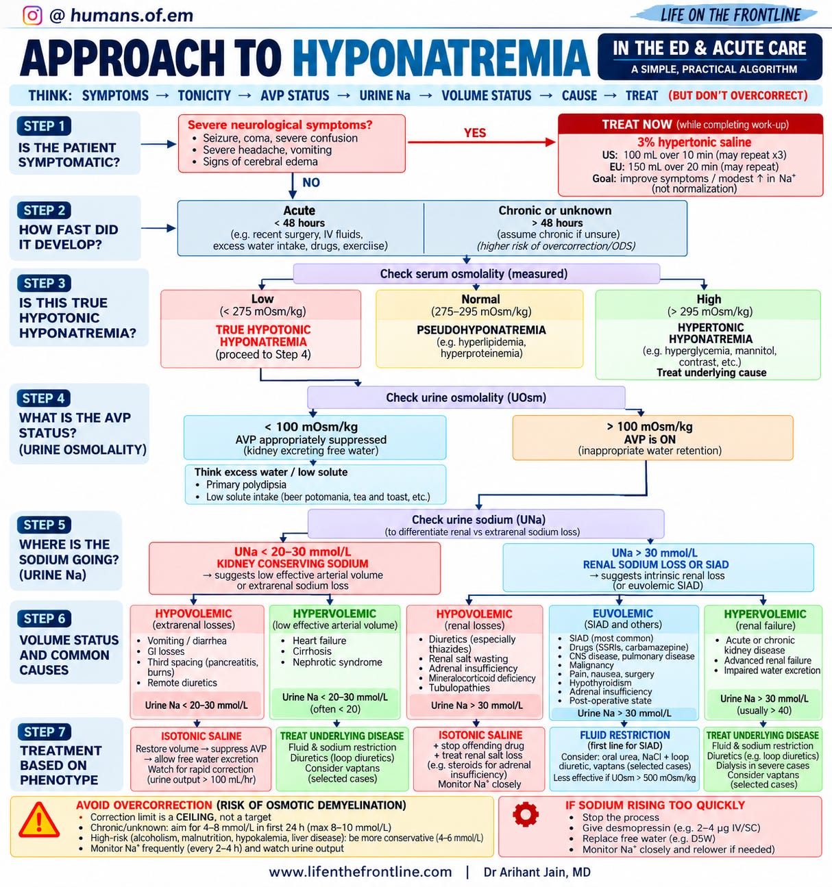
Now comes the most important part: Treatment
There is no single treatment for “hyponatremia.”
Treatment depends on why the sodium is low and whether the patient is symptomatic.
1. Hypertonic saline: treat symptomatic cerebral edema
For acute or severe symptomatic hyponatremia, 3% saline is the treatment of choice.
The review describes bolus-based approaches from both US and European recommendations.
US approach
100 mL of 3% saline over 10 minutes
May be repeated, with a maximum of three boluses.
European approach
150 mL of 3% saline over 20 minutes
Again, repeat dosing may be required according to clinical and biochemical response.
The important concept is:
Give enough hypertonic saline to improve the neurological emergency — not enough to normalize the sodium.
2. Isotonic saline: treat hypovolemic hyponatremia
If the problem is volume depletion:
Restore circulating volume.
Giving isotonic saline reduces the non-osmotic stimulus for AVP release.
As AVP falls, the kidney can suddenly begin excreting large amounts of free water.
And this creates an important ED trap:
The sodium can suddenly rise much faster than expected.
An increase in urine output—particularly >100 mL/hour—can be an early warning sign of impending overcorrection.
So after starting fluids:
Don’t just watch the sodium.
Watch the urine output.
3. Fluid restriction: mainly for SIAD
For patients with SIAD without severe neurological symptoms, fluid restriction is commonly used.
But its effectiveness depends on how strongly AVP is driving water retention.
A very concentrated urine—for example, UOsm >500 mOsm/kg—predicts a poorer response to fluid restriction.
So:
Fluid restriction is not automatically effective just because the patient has SIAD.
4. Other options
Depending on the underlying cause and clinical setting, other approaches may include:
SIAD
Fluid restriction
Oral urea
Loop diuretic + oral sodium in selected patients
Vasopressin receptor antagonists in selected circumstances
Hypervolemic hyponatremia
Treat the underlying disease
Fluid/sodium restriction where appropriate
Diuretics
Selected use of vasopressin antagonists
The evidence and guideline recommendations differ for some of these therapies, particularly vaptans and loop-diuretic strategies.
And finally: Don’t forget overcorrection
This may be the most important safety principle in managing chronic or unknown-duration hyponatremia.
The correction limit is a ceiling — not a target.
For chronic or unknown-duration hyponatremia, the review summarizes:
European guidance: maximum 10 mmol/L in the first 24 h, then 8 mmol/L/day
US recommendations: generally a more conservative target of 4–8 mmol/L/day, with a maximum limit of 8–12 mmol/L/day
In patients at high risk of ODS—such as those with alcoholism, malnutrition, hypokalemia or advanced liver disease—more conservative correction is advised.
The exact threshold should therefore be interpreted in the context of chronicity and ODS risk.
What if the sodium is rising too quickly?
Stop the process.
Then:
Replace free-water losses
Desmopressin
±
5% dextrose / free water
The combination of desmopressin and free-water replacement can be used to stop further sodium rise or, when necessary, actively relower the sodium toward the intended target.
This is particularly important when the patient suddenly begins producing large volumes of dilute urine after the underlying stimulus for AVP secretion disappears.
One final principle
Hyponatremia is not simply a problem of “too little sodium.”
It is usually a problem of too much water relative to sodium.
Once you start thinking in terms of:
Symptoms → Osmolality → AVP → Urine Osm → Urine Na → Cause → Treatment
the seemingly complicated hyponatremic patient becomes much easier to approach.
Treat the brain.
Define the physiology.
Find the cause.
Correct carefully.
That is the bedside approach to hyponatremia.
DISCLAIMER: For Educational Purposes only!