Oxygen-carrying capacity → PaO₂, Hb, total Hb and dyshemoglobins
Electrolytes, Hb, Hct, Osmolality, etc.
It can also be used to assess response to interventions such as oxygen therapy and to monitor the severity or progression of disease.
2. Getting the sample right
Before puncture
Allen’s test as a prerequisite
Slight wrist extension for radial artery sampling
Clean the site with 70% alcohol
Use an appropriate heparinised syringe
Enter the skin at approximately 45° for radial artery sampling
Obtain approximately 1 mL of blood
Do not pull back the syringe plunger
If an air bubble enters the syringe:
Remove it immediately.
Then apply firm pressure to the puncture site for at least 5 minutes.
3. The sample can be wrong even when the puncture was right
Several pre-analytical errors can alter the ABG:
Too much or too little anticoagulant
The source highlights the potential effects of excess/inadequate anticoagulant, including electrolyte binding.
Venous contamination
Mixing venous and arterial blood during puncture can produce misleading values.
Air bubbles
Air exposure can:
↑ PaO₂ ↓ PaCO₂
Inadequate mixing
Can cause RBC stacking and compromise the sample.
4. Time matters
Analyse within 5 minutes Maximum: 15 minutes
If a delay of >30 minutes is expected, it is recommended to keep in a glass syringe with an ice slurry, with iced-sample analysis possible for up to approximately 1 hour.
Practical takeaway:
Don’t let a good arterial puncture become a bad ABG because of poor handling.
5. When should you avoid a particular puncture site?
These cautions:
Negative Allen’s test
Surgical shunt/fistula or bypass graft
Infection at the site
Limb ischemia/Raynaud’s disease
Bleeding diathesis — described as a relative contraindication in the post-thrombolysis setting
6. Complications of ABG sampling
Even a routine ABG is an arterial puncture. Potential complications include:
Arterial spasm Hematoma Arterial occlusion Air/thrombus embolism Local infection Nerve or vessel injury Vasovagal response Needle-stick injury
7. Now comes the part most people skip…
CHECK THE ABG BEFORE INTERPRETING IT
Before jumping to:
❌ “It’s metabolic acidosis.” ❌ “It’s respiratory alkalosis.” ❌ “The patient is hypoxic.”
First ask:
Does the ABG make physiological sense?
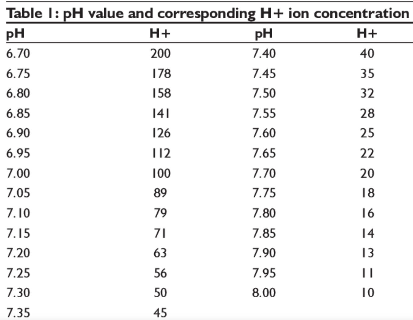
The modified Henderson–Hasselbalch relationship
[H⁺] = 24 × PaCO₂ / HCO₃⁻
Use the reported PaCO₂ and HCO₃⁻ to estimate the corresponding hydrogen ion concentration.
Then compare it with the reported pH.
If the pH and calculated H⁺ are inconsistent:
Question the validity of the ABG.
A corresponding pH–H⁺ table as a bedside reference.
The Frontline Takeaway
Don’t interpret the numbers before validating the sample.
Good ABG interpretation begins before the ABG reaches the analyser.
Collect correctly → Handle correctly → Process promptly → Check consistency → Then interpret.
Coming next…
ABG series #2 — Oxygenation
Hypoxemia ≠ Hypoxia
We’ll start with:
PaO₂ → SaO₂ → FiO₂ → P/F ratio → A–a gradient
And then ask the more important question:
Why is this patient hypoxemic?
Follow @humans.of.em for the next part of the ABG series.